ECG Challenge

This is an ECG of a 65 years old male. He had a device implanted 1 week back. Chances are, if you are not well acquainted with ECGs of Pacemakers you'll miss the pacemaker 'spikes', and what information these spikes present. In this ECG, there is an obvious left axis deviation and 'Poor progression' of R wave in leads V1 to V6. If you look closely ,however, before every QRS complex, you can notice a vertical line known as the 'Pacemaker spike'.
 |
This Pacemaker spike appears from the current that is delivered by the Pacemaker and subsequently a QRS complex forms after that, if the current delivered is in the ventricles. Now, an Atrial lead also does the same when producing a current, but here we notice that the P waves are not preceded by any 'spike'. However, every P wave is followed by a spike and then a QRS complex. This leads us to the believe that the Pacemaker is sensing native P waves through the Atrial lead in the Right Atrium (where it is always placed) and Pacing through the ventricular lead (A Sense V Pace). Although a closer inspection reveals something else.
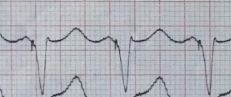 |
| Each P wave followed by spike and then QRS |

So we have been able to establish 3 things from the ECG so far; First that it is a Paced rhythm, Second it is a Dual Chamber Pacemaker and Third that it is Pacing the Atria and Ventricles.
Wait, but there is one more aspect. You see, the spikes before the ventricles are actually 2 separate spikes. This means that the ventricles are being stimulated at 2 different places. In fact, it is a CRT (Cardiac Synchronization Therapy) device. Here there are 3 leads in the heart, One in the Right Atrium, One in the Right Ventricle, and a 3rd one inserted through the Right Atrium into the Coronory Sinus all the way into the Epicardial veins outside the Left Ventricle and as a result it is stimulating the Left Ventricle as well as the Right Ventricle to improve the Ejection Fraction.
 |
| 2 different spkies in CRT device |


Comments
Post a Comment