ECG Challenge 3 - Inverted waves
This is an ECG of a 55 years old Male with no known co-morbids. He presented in Emergency with history of palpitations.
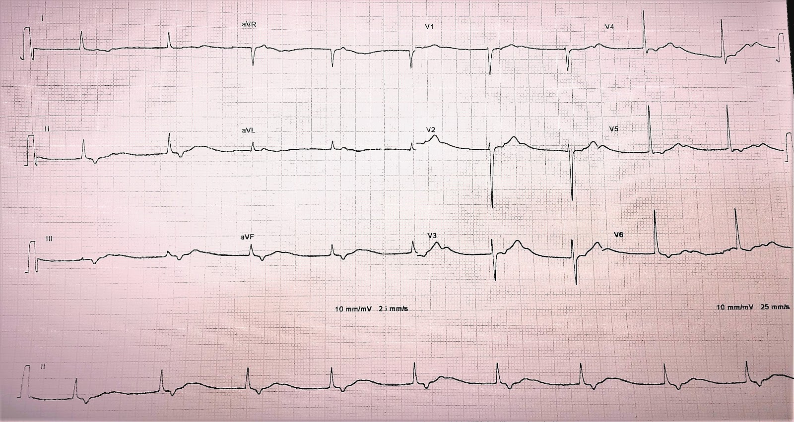
So, what is your opinion about this ECG? Take your time, have a detailed look at it and when you are ready, proceed further for a detailed analysis.
First of all, as discussed in the previous case, there are no P waves before the QRS complexes. This means that the Sinus node is not functioning properly. Lets zoom in an have a look:

The underlined part is where a normal P wave should be and we don't see any here. But what about those inverted waves just after the QRS complex?

Highlighted here by red arrows, are these inverted T waves or is it something else?
If we look closely at the Precordial leads especially V2 and V3, we can see that these little humps are actually not T Waves but the T waves come after these. Have a look:

The T waves are those that appear here in blue circles, those in green circle are actually U waves. So what about the waves we are talking about, those encircled here in red. Well Those are actually P waves; Retrograde P Waves!
This is Nodal Rhythm, most likely arising due to Sinus Arrest; The sinus node is not generating any activity and as a result the AV Node has started generating its own rhythm. The current moving in ventricles gives the QRS complex and that moving upward into the atria produces these inverted P waves.
But how do we know it is from the AV node and not the Ventricles? Well, that's because the QRS generated is Narrow (<120ms), Rhythm arising from the ventricles is almost always broad complexed.
Therefore, this is a case of Sinus Arrest with Nodal Rhythm,
Your input is valuable, please comment below to let us know what do you think about this ECG.

Comments
Post a Comment